Decoding ultrasound during pregnancy – tables of norms screening ultrasound of a pregnant woman
An ultrasound scan is an opportunity to find out about the state of health of a child while he is in the womb. During this study, the expectant mother for the first time hears her child’s heart beating, sees his arms, legs, and face. If desired, the doctor can provide the sex of the child. After the procedure, the woman is given a conclusion, in which there are quite a few different indicators. It is in them that we will help you figure it out today.

The content of the article:
Norms of ultrasound results of a pregnant woman in the first trimester
A pregnant woman does her first ultrasound screening at 10-14 weeks of pregnancy. The main objective of this study is to find out if this pregnancy is ectopic.
In addition, special attention is paid to the thickness of the collar zone and the length of the nasal bone. The following indicators are considered within the normal range – up to 2.5 and 4.5 mm, respectively. Any deviations from the norms can be a reason for visiting a geneticist, as this may indicate various defects in the development of the fetus (Down, Patau, Edwards, Triplodia and Turner syndromes).
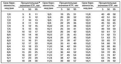
Also, during the first screening, the coccygeal-parietal size is assessed (norm 42-59 mm). However, if your numbers are slightly abnormal, don’t panic right away. Remember that your baby is growing daily, so the numbers at 12 and 14 weeks will differ significantly from each other.

Also, during an ultrasound scan, the following are assessed:
- Baby’s heart rate;
- Umbilical cord length;
- The state of the placenta;
- The number of vessels in the umbilical cord;
- Placenta attachment site;
- Lack of dilatation of the cervix;
- Absence or presence of a yolk sac;
- The appendages of the uterus are examined for the presence of various anomalies, etc.

After the end of the procedure, the doctor will give you his opinion, in which you can see the following abbreviations:
- Coccyx-parietal size – CTE;
- Amniotic index – AI;
- Biparietal size (between the temporal bones) – BPD or BPHP;
- Frontal-occipital size – LZR;
- The diameter of the ovum is DPR.

Decoding of ultrasound of the 2nd trimester at 20-24 weeks of pregnancy
The second ultrasound screening the pregnant woman should undergo at a period of 20-24 weeks. This period was not chosen by chance – after all, your baby has already grown up, and all his vital systems have been formed. The main purpose of this diagnosis is to identify whether the fetus has malformations of organs and systems, chromosomal pathologies. If developmental deviations that are incompatible with life are identified, the doctor may recommend an abortion, if the terms still allow.

During the second ultrasound, the doctor examines the following indicators:
- Anatomy of all internal organs of the baby: heart, brain, lungs, kidneys, stomach;
- Heart rate;
- Correct structure of facial structures;
- Fetal weight, calculated using a special formula and compared with the first screening;
- The state of the amniotic fluid;
- The state and maturity of the placenta;
- The gender of the child;
- Single or multiple pregnancy.

At the end of the procedure, the doctor will give you his opinion on the condition of the fetus, the presence or absence of developmental defects.
There you can see the following abbreviations:
- Abdominal circumference – coolant;
- Head circumference – OG;
- Frontal-occipital size – LZR;
- Cerebellum size – RM;
- Heart size – RS;
- Thigh length – DB;
- Shoulder length – DP;
- Chest diameter – DGrK.

Decoding ultrasound screening in the 3rd trimester at 32-34 weeks of pregnancy
If the pregnancy was proceeding normally, then the last ultrasound screening is performed at 32-34 weeks.
During the procedure, the doctor will assess:
- all fetometric indicators (DB, DP, BPR, OG, coolant, etc.);
- the condition of all organs and the absence of malformations in them;
- presentation of the fetus (pelvic, head, transverse, unstable, oblique);
- state and place of attachment of the placenta;
- the presence or absence of an umbilical cord entanglement;
- well-being and activity of the baby.

In some cases, the doctor prescribes another ultrasound scan before childbirth – but this is more the exception than the rule, because the baby’s condition can be assessed using cardiotocography.
Remember – the doctor should decipher the ultrasound, taking into account a large number of different indicators: the condition of the pregnant woman, the features of the parents’ constructions, etc.

Each child is individual, so he may not correspond to all the average indicators.
All information in this article is for educational purposes only. The сolady.ru website reminds you that you should never delay or ignore your visit to a doctor!
What to give a friend?
Gift Certificate! You can give it to your loved one or use it yourself.
And we also give away a certificate for 3000 rubles every month. among new email subscribers. Subscribe!
Select a certificate in the store
Visit Bologny for more useful and informative articles!


